Malignant Mesothelioma Ct Radiology - Clinical Staging Of Malignant Pleural Mesothelioma Current Perspectiv Lctt : mesothelioma affects the mesothelium, or thin linings covering organs, such as the lungs (pleural mesothelioma) and abdomen (peritoneal mesothelioma).
Malignant Mesothelioma Ct Radiology - Clinical Staging Of Malignant Pleural Mesothelioma Current Perspectiv Lctt : mesothelioma affects the mesothelium, or thin linings covering organs, such as the lungs (pleural mesothelioma) and abdomen (peritoneal mesothelioma).. Multidetector ct findings and differential diagnoses of malignant pleural mesothelioma and metastatic pleural diseases in korea. However, tumor invasion into the chest wall or diaphragm can be underestimated by ct. Role of ct, mri, and pet/ct in staging evaluation and treatment considerations. The ct scans of 84 patients with proved malignant pleural mesothelioma were retrospectively evaluated. This is a case of pathology proven metastatic malignant mesothelioma.
mesothelioma radiology benign or malignant. This is a case of pathology proven metastatic malignant mesothelioma. ct scans are preferred for staging tumors and are vital for patients with malignant pleural mesothelioma, yendamuri explained. This is an uncommon benign primary peritoneal tumor that has no relation with the malignant mesothelioma. Nickell lt jr et al:
Malignant Pleural Mesothelioma Evaluation With Ct Mr Imaging And Pet Radiographics from pubs.rsna.org malignant pleural mesothelioma (mpm) is a rare malignant neoplasm of the pleura that typically affects individuals occupationally exposed to asbestos through a variety of industries. About cancer cancer research uk. Mirvis s, dutcher jp, haney pj, et al. Symptoms or abnormalities not consistent with the initial. Fifty percent of these tumors tend to locally recur after surgical resection. mesothelioma radiology benign or malignant. • pleural plaques are present in approximately 20% of mesothelioma patients. ct of malignant pleural mesothelioma.
Computed tomography (ct) enables early detection of small pleural tumors and pleural effusions and definition of the extension of the tumor along the pleural surfaces and fissures.
Multidetector ct findings and differential diagnoses of malignant pleural mesothelioma and metastatic pleural diseases in korea. Magnetic resonans imaging (mri) is useful for detection of extension of disease, especially to the chest wall and diaphragm (moore et al., 2008, wang et al., 2004). J comput assist tomogr 1983; malignant pleural mesothelioma (mpm) is an aggressive thoracic malignancy with a dismal prognosis. 10 the british thoracic society (bts) statement on malignant mesothelioma specifically recommends scanning at 60 s delay to achieve a. In both cases, a histopathological examination of the pleura confirmed the diagnosis of epithelioid malignant. malignant pleural mesothelioma (mpm) is traditionally characterized by local destructive spread of the pleura and surrounding tissues. Symptoms or abnormalities not consistent with the initial. These findings are suggestive of scrotal mesothelioma. This is an uncommon benign primary peritoneal tumor that has no relation with the malignant mesothelioma. About cancer cancer research uk. Use of ct for tumor staging. However, tumor invasion into the chest wall or diaphragm can be underestimated by ct.
This review is based on a presentation given by angela levy and adapted for the radiology assistant by robin smithuis. The radiology of thoracic malignant mesothelioma. Alexander e, clark ra, colley dp, mitchell se. mesothelioma, peritoneal angiography computed tomography introduction reports of computed tomography (ct) in mesothelioma describe peritoneal involvement that may be extensive, with confluent tumor in layers, masses, and/or nodules and mesenteric infiltration 1, 2. It can have a myxoid stroma resulting in a low attenuation on ct and a high attenuation on t2wi.
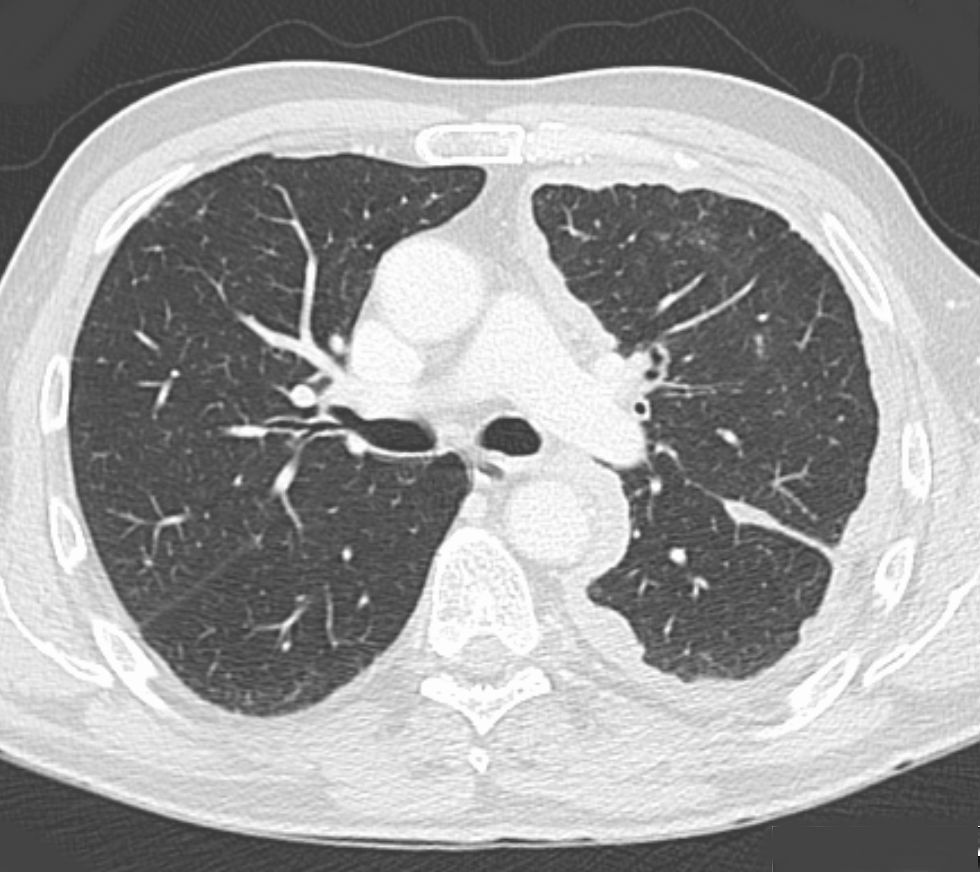
A 71 Year Old Patient With Pleural Mesothelioma Ct Scan Top A Download Scientific Diagram from www.researchgate.net Your email address will not be published. Pleural biopsies in patients with suspected malignant pleural mesothelioma (mpm) are often inconclusive resulting in repeat diagnostic procedures. These findings are suggestive of scrotal mesothelioma. An educational website focused on the intersection of nuclear medicine and radiology. We show the ct and mri images of the tumor we experienced. This is a case of pathology proven metastatic malignant mesothelioma. Nowak ak, francis rj, phillips mj, et al. Kitajima k, doi h, kuribayashi k, et al.
mesothelioma is an uncommon malignant tumor that arises from mesenchymal tissue and involves pleura, peritoneum, pericardium and very rarely, the tunica vaginalis (<
mesothelioma radiology benign or malignant. Review article comparative interpretation of ct and standard radiography of the pleura. At ct, the soft tissue density of tumor tissue can be readily distinguished from the adjacent pleural effusion, but the nodules may on occasion be so. Your email address will not be published. Superior vena cava syndrome with malignant reasons nejm. malignant mesothelioma is a type of cancer that affects the tissue that lines your lungs, heart, stomach, and other organs. Okten f, köksal d, onal m, ozcan a, sims¸ek c, ertürk h. Although the chest film findings of pleural mesothelioma are well described, there are few descriptions of the findings of computed tomography (ct). There are different types of mesothelioma, including pleural and peritoneal mesothelioma. The exact prevalence is unknown but it is estimated that mesotheliomas represent less than 1% of all cancers. Use of ct for tumor staging. Older patients with early stage mpm are more likely to. In this retrospective study, we reviewed a cohort of 164 mpm patients referred to a phase i trials unit, aiming to describe identified metastatic sites, and correlate with clinical.
An educational website focused on the intersection of nuclear medicine and radiology. Wechsler rj, rao vm, steiner rm. Alexander e, clark ra, colley dp, mitchell se. Indeed, ct still presents some intrinsic limitations such as a poor contrast The results are malignant epitheloid cell forms a cohesive nest, glandular structure and a lot of micropapillae in accordance with malignant mesothelioma (epitheloid mesothelioma).
Malignant Pleural Mesothelioma Pacs Suche Fur Radiologen from upload.wikimedia.org ct in differential diagnosis of diffuse pleural. mesothelioma, peritoneal angiography computed tomography introduction reports of computed tomography (ct) in mesothelioma describe peritoneal involvement that may be extensive, with confluent tumor in layers, masses, and/or nodules and mesenteric infiltration 1, 2. The ct scans of 84 patients with proved malignant pleural mesothelioma were retrospectively evaluated. mesothelioma has a long latency period of 20 to 40 years, and many patients do not have symptoms until the disease is in its later stages, when metastasis is more likely to occur. mesothelioma radiology benign or malignant. Twenty patients (24%) had been exposed to erionite and 64 patients (76%) had been exposed to. This review is based on a presentation given by angela levy and adapted for the radiology assistant by robin smithuis. Mirvis s, dutcher jp, haney pj, et al.
There are different types of mesothelioma, including pleural and peritoneal mesothelioma.
Asbestos plaques are not a… This is an uncommon benign primary peritoneal tumor that has no relation with the malignant mesothelioma. ct of malignant pleural mesothelioma. malignant mesothelioma is a type of cancer that affects the tissue that lines your lungs, heart, stomach, and other organs. malignant mesothelioma is a rare and aggressive cancer. Magnetic resonans imaging (mri) is useful for detection of extension of disease, especially to the chest wall and diaphragm (moore et al., 2008, wang et al., 2004). There's no history of syncope or. We investigated pleural cle imaging as a biopsy guidance technique to distinguish malignant from benign pleural disease. In both cases, a histopathological examination of the pleura confirmed the diagnosis of epithelioid malignant. It can have a myxoid stroma resulting in a low attenuation on ct and a high attenuation on t2wi. Indeed, ct still presents some intrinsic limitations such as a poor contrast About cancer cancer research uk. In each case the ct showed.


0 Comments